FIGURE 9.
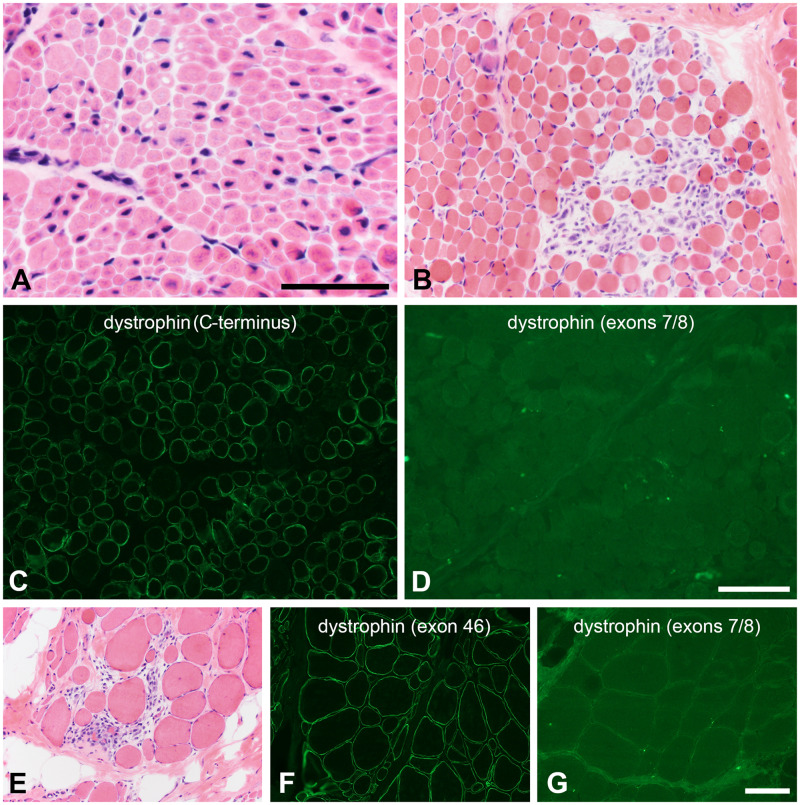
Resolving variants of unknown significance (VUS). (A) The histopathologic phenotype of X-linked myotubular myopathy is sufficiently specific that a VUS in MTM1 may be resolved by simply evaluating a hematoxylin and eosin-stained cryosection. In the patient illustrated here, a small in-frame deletion in the MTM1 gene was classified a VUS and the muscle biopsy revealed central nuclei, central vacuoles, central consolidation of sarcoplasmic organelles, and numerous, abnormally small muscle fibers. Scale bar: 50 µm. (B–D) After identifying a VUS in DMD (c.79G>C, p. A27P), this 2-year-old boy underwent a muscle biopsy to clarify the pathogenicity of the variant. The histopathology is dystrophic (B). Dystrophin immunofluorescence intensity is mildly to moderately reduced using antibodies directed at the carboxy terminus and rod domains; the C-terminus is shown (C). No staining was observed with an actin binding domain antibody directed at an epitope in exons 7/8 near the amino terminus (D). (E–G) After identifying a VUS in DMD (c.76A>C, p. N26H), this 11-year-old boy underwent a muscle biopsy to clarify the pathogenicity of the variant. The histopathology is dystrophic (E). Dystrophin immunofluorescence intensity is mildly to moderately reduced using antibodies directed at the carboxy terminus and rod domains; exon 46 is shown here (F). No staining was observed with an actin binding domain antibody directed at an epitope in exons 7/8 near the amino terminus (G). The scale bar in panel D is 100 µm for panels B–D. The scale bar in panel G is 100 µm for panels E–G.

