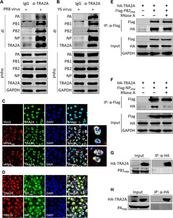
Fig. 1. TRA2A associates with vRNP.

(A and B) A549 cells were infected with either the YS (A) or the PR8 (B) virus at a multiplicity of infection (MOI) of 0.01 for 24 hours; cell lysates were immunoprecipitated with an anti-TRA2A antibody or the control immunoglobulin G (IgG) and then analyzed by Western blotting. GAPDH, glyceraldehyde-3-phosphate dehydrogenase. (C) A549 cells were infected with the YS or PR8 virus at an MOI of 5. At 4 hours post-infection (hpi), cells were subjected to RNA FISH combined with immunofluorescence to detect M vRNA and TRA2A protein. Scale bars, 10 μm. DAPI, 4′,6′-diamidino-2-phenylindole. (D) A549 cells were infected with either YS or PR8 virus at an MOI of 0.01. At 24 hpi, cells were fixed and analyzed for the colocalization of TRA2A with NP. Scale bars, 20 μm. (E and F) Human embryonic kidney (HEK) 293T cells were transfected with the indicated plasmids for 24 hours. The cell lysates were treated with or without 100 U of ribonuclease A (RNase A) at 37°C for 1 hour. CoIP assay was performed using an anti-Flag antibody and analyzed by Western blotting. (G and H) HEK 293T cells were transfected with the indicated plasmids for 24 hours. Cell lysates were immunoprecipitated with an anti-hemagglutinin (HA) antibody and then analyzed by Western blotting.
