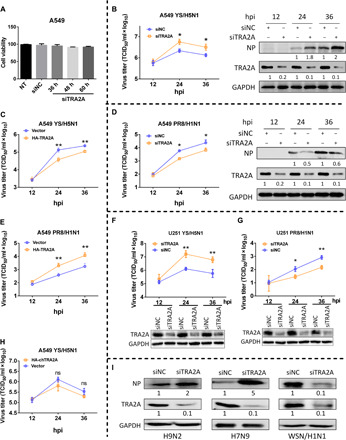
Fig. 2. TRA2A inhibits YS replication but enhances PR8 replication.

(A) A549 cells were seeded on the 96-well plates, and cell viability was detected by Cell Counting Kit-8 assay at 36, 48, and 60 hours after transfection. NT, non-treated. (B to H) A549 (B to E and H) or U251 (F and G) cells were transfected with either HA-TRA2A, HA-chTRA2A, or pCAGGS vector or either siNC (negative control) or siTRA2A for 24 hours and then infected with the YS (B, C, F, and H) or PR8 (D, E, and G) virus at an MOI of 0.01. Cell culture supernatants were collected at 12, 24, and 36 hpi. Virus titers were determined by TCID50 assay on MDCK (Madin-Darby canine kidney) cells. A549 cell lysates were analyzed by Western blotting, and the silence efficiency of TRA2A and changes of NP were quantified by ImageJ and normalized to GAPDH (C and E). (I) A549 cells were transfected with either siNC or siTRA2A for 24 hours and infected with an avian virus H9N2 or H7N9 or a human virus WSN/H1N1 strain at an MOI of 0.01. Cells lysates were subjected to Western blotting analysis. GAPDH was used as loading control. Each protein band was quantified by ImageJ and normalized to GAPDH levels. Means ± SD (error bars) of three independent experiments are indicated (*P < 0.05 and **P < 0.01).
