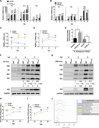
Fig. 5. M segment 334 site single mutation alters its mRNA splicing and virus replication.

(A to D, F, and G) A549 cells were infected with either the PR8-WT, PR8-M334C, YS-WT, or YS-M334C virus at an MOI of 5; the total RNA was isolated and analyzed by RT-qPCR assay (A and B). Ratios of M2/M1 mRNA are presented (C and D). Cells lysates were subjected to Western blotting analysis using antibodies against respective influenza virus proteins. Each protein band was quantified by ImageJ and normalized to GAPDH levels (F and G). (E) A549 cells were infected with the WT or mutated YS (right) or PR8 (left) virus (MOI, 1) for 12 hours, and the mRNAs were purified with either the control IgG or the TRA2A antibody and analyzed by RT-qPCR assay. (H and I) Growth curves of the indicated virus. (J) A549 cells were transfected with siNC or siTRA2A for 24 hours and then infected with the indicated virus for 6 hours. Cells were stained with rabbit anti-HA antibody followed by fluorescein isothiocyanate (FITC)–goat anti-rabbit IgG and analyzed by flow cytometry. The data represent means ± SD (error bars) of three independent biological replicates (ns, not significant; *P < 0.05, **P < 0.01, and ***P < 0.001).
