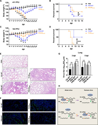
Fig. 6. Pathogenicity of WT YS or PR8 and their mutated viruses at 334 site in mice.

Six-week-old female BALB/c mice were intranasally inoculated with the indicated doses of the PR8-WT, PR8-M334G, YS-WT, or YS-M334C virus or the phosphate-buffered saline (PBS) control. (A to D) Weight loss and mortality of mice infected with each indicted virus. Body weight of the WT and 334 mutant groups were compared and statistically analyzed. Error bars represent means ± SEM (n = 10). Statistical analysis was used by two-tailed analysis of variance (ANOVA) with Bonferroni post test. PFU, plaque-forming units. (E) Pathological lesions in the lungs of mice infected with the indicated virus at 3 dpi with hematoxylin and eosin (H&E) staining. Scale bars, 100 μm. (F) Immunofluorescent staining of lung sections of mice infected with the indicated virus at 3 dpi. The viral NP antigen was stained green, and the nucleus was stained blue. Scale bars, 50 μm. (G) Virus titers in the lungs of infected mice (n = 3) at 3 dpi (left) and 5 dpi (right). Error bars represent means ± SD. Statistical analysis was performed by using one-tailed method. EID50, 50% egg infectious dose. (H) Model for avian IAV overcomes the huTRA2A host barrier. *P < 0.05, **P < 0.01, and ***P < 0.001.
