
Figure 1.
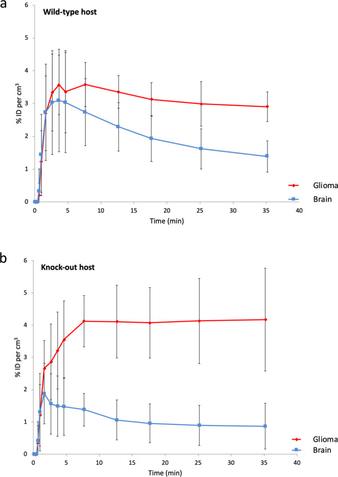
Tracer kinetic curves showing the behaviour of the ligand PBR111 in implanted TSPO expressing glioma (red), compared to the ligand kinetics of PBR111 in the tissue of the surrounding brain (blue). (a) Wild-type host: The kinetics of [18 F]PBR111 demonstrate the increased retention of [18 F]PBR111 in the TSPO expressing syngeneic tumour (red), while healthy, normal cerebral tissue (blue) (excluding cerebellum) in Tspo+/+ animals has low to absent [18 F]PBR111 retention. ID = injected dose; n = 3; error bars denote standard deviation. (b) Knock-out host: Tspo−/− mice do not have any specific binding of [18 F]PBR111 in any organ. ID = injected dose; n = 3; error bars denote standard deviation.