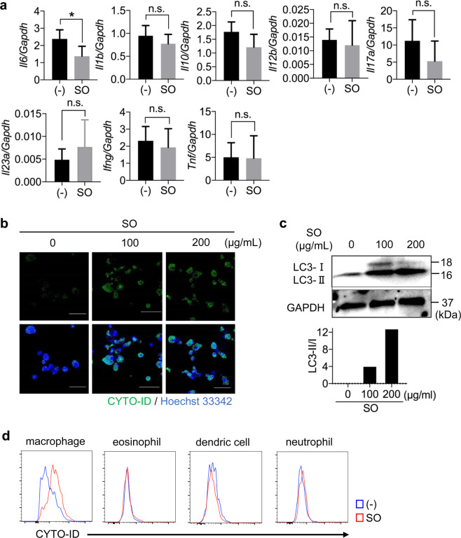
Figure 4.
SO induced autophagy in bone marrow-derived Mφ (BMMφ) and intestinal Mφ. (a) Expression of the indicated genes in colonic lamina propria cells from mice treated with or without SO in the steady state. Data are mean ± SD of three independent experiments. Data were analyzed by unpaired t-test, *p < 0.05. n.s., not significant. (b) CYTO-ID staining in BMMφ treated with or without SO for 24 hours. (c) BMMφ were cultured in the presence or absence of SO for 24 hours. The immunoblotting was performed with the indicated antibodies. The graphs show the ratio of LC3-II to LC3-I. Full-length blots are shown in Supplementary Fig. S10c. (d) C57BL/6J mice administered 2% DSS with or without SO in drinking water for 7 days and colonic lamina propria cells were isolated. The autophagosome formation stained by CYTO-ID in macrophages, eosinophils, dendric cells and neutrophils were analyzed with flow cytometry. Data are representative of two independent experiments.

