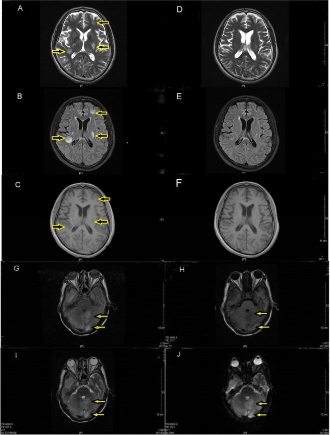
Figure 2.
Supratentorial lesions as showed in picture A, B, C, D; Picture A, B, C are T1W1 imaging, T2W1 and Flair imaging respectively of a patient diagnosed with CM before being treated with GCs, 43th days after being admitted to hospital. T1W1 displayed low signal while T2W2 and Flair imaging displayed high signal which distributed beneath the left frontal cortex, within the white matter of basal ganglia and the right cistern (as indicated by arrows). Picture D, E, F are the same T1W1 imaging, T2W2 and Flair imaging of the patient after being treated with GCs for 21 days. No significant abnormality was seen in T1W1 and T2W2 imaging while the high signal in the left frontal cortex and the white matter of right cistern showed by Flair was obviously decreased or even disappeared in comparison to the previous (relatively compare with the area indicated by arrows in Picture A, B, C). Infratentorial lesions showed in picture G, H, I, J by Flair, T1W1, T1W2 and DWI imaging, the yellow arrow represents inflammatory signal. In this figure they weren’t the same person for pictures of supratentorial and infratentorial lesions.

