Figure 1.
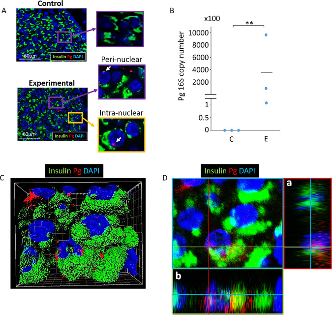
Pg/gingipain translocates to the pancreas and is present in β-cells. (A) Representative result using IF microscopy showing Pg/gingipain in pancreatic islets in experimental but not control animals. White arrows point to Pg/gingipain. Scale bar represents 60μm. N = 9 mice/experimental group and N = 10 mice/control group. (B) Results from qPCR for Pg 16 S rRNA genes performed on formalin fixed paraffin embedded (FFPE) sections (5/animal) show that Pg is present in islets from experimental animals but not controls. N = 3 mice/group. The difference between groups is statistically significant (p** < 0.01). (C) Representative 3-D image from confocal microscopy of mouse pancreatic islet showing that Pg is localized in β-cells. (D) Orthogonal analysis of image confirming intra-nuclear localization of Pg in β-cells. a & b are orthogonal projections showing the side views of the main image cut through along vertical (red) and horizontal (green) lines. Green: insulin, Red: Pg, Blue: nuclei.

