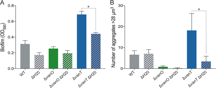
Fig. 5. Biofilm formation and aggregation of V. anguillarum 90-11-287 is enhanced at low-cell density and affected by the H20-like prophage.
(a) Quantification of biofilm formation after 48 h of incubation. (b) Aggregates with a biovolume larger than 28 µm3 based on CLSM z-stack images in an area of 159.73 × 159.73 µm. An asterisk indicates a significant difference between the prophage-harboring and prophage-free mutants (ANOVA and Tukey’s multiple comparison test, α = 0.05) and error bars represent standard error of the mean (N = 3 for crystal violet assays and N = 4 for CLSM quantification).

