Fig. 2.
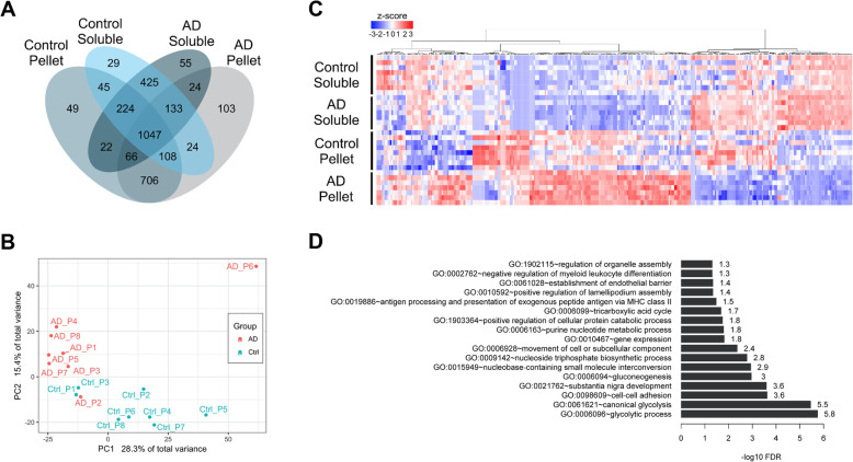
AD and control brains segregate into distinct clusters. Bioinformatic analysis of aggregated proteins in AD and control brains. a Venn diagram of expressed proteins (more than half of the biological replicates with non-zero counts) in soluble and pellet fractions of AD and control individuals. b Principle component analysis (PCA) plot of insoluble ratios for all proteins across individuals. Red = AD patients, blue = control patients. c Heatmap of 342 proteins with significantly different insoluble ratios between AD and control individuals. Values are z-scaled quantile normalized log-transformed spectral counts. Red = high, blue = low. d Bar plot of significantly enriched gene ontology (GO) biological process (BP) terms for the 342 proteins

