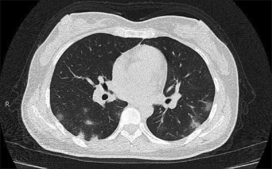
Figure 2.

Axial noncontrast image of a computed tomography of the chest performed 5 days after the onset of symptoms shows patchy, peripheral predominantly nodular areas of consolidation and ground-glass opacities. Zhang W, Shi H. Evolving COVID-19 Pneumonia. Radiology Case Collection doi: 10.1148/ cases. 20201558. Published online March 17, 2020. ©Radiological Society of North America (Reproduced with permission from the Radiological Society of North America)
