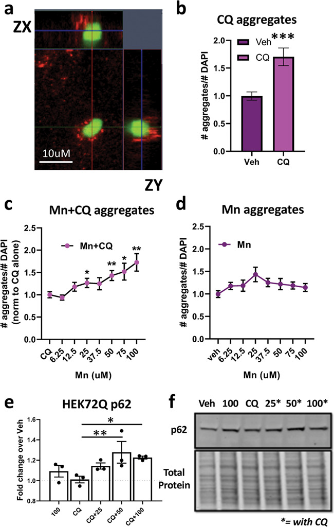
Figure 6.

Mn increases the association of autophagosomes with mutant HTT aggregates. (a) Representative 63× confocal image of GFP-HTT-72Q aggregates (green) near LC3 puncta (red) in transfected HEK293 cells after treatment with 75 uM Mn + 10 uM CQ. Includes ZX and ZY view. (b) Quantification of relative number of aggregates after CQ (10 uM) treatment for 72 h. Asterisks, significant difference by student’s t-test. (c) Quantification of number of aggregates after 72 h treatment with increasing amounts of Mn + 10 uM CQ. Univariate ANOVA; F(4.085, 98.04) = 5.552; P = 0.0004. (d) Quantification of number of aggregates after 72 h treatment with increasing amounts of Mn alone. Univariate ANOVA; F(4.046, 89.02) = 1.369; P = 0.2907. Quantification of (c) is normalized to CQ alone and quantification of (d) is normalized to no treatment. N = 3 biological replicates, each with 10 images. (e) Quantification of p62 after 72 h treatment with Mn (25/50/100 uM) and/or CQ (10 uM), in 72Q-GFP HEK293 cells. N = 3. Univariate ANOVA; F (4, 8) = 5.743; P = 0.0177. (f) Representative western blot for HEK72Q p62. Asterisks, exposed with 10 uM CQ. Error bars = SEM; *P < 0.05, **P < 0.01, ***P < 0.001. Asterisks, significance difference by Dunnet’s multiple comparison tests.
