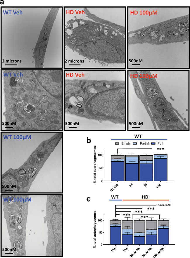
Figure 8.

Acute Mn exposure attenuates empty APV phenotype in Q111 STHdh cells. (a) Representative transmission electron microscopy (TEM) images of STHdh WT Q7/Q7 (blue) and HD Q111/Q111 (red) cells with no treatment (Veh) or 100 uM Mn for 24 h. (b and c) Quantification of empty, partial or full autophagosomes after 24 h treatment with 25/50/100 uM Mn in STHdh WT Q7/Q7 (b) and HD Q111/Q111 (c) cells. Note: for panel c, vehicle-treated WT is re-plotted on the left for comparison. A total of 15–20 images per condition were analyzed across two independent TEM sections from two independent biologically experimental sets giving a technical n = ~ 30–40 per condition. Error bars = SEM; Asterisks, significant difference by chi-square analysis. *P < 0.05, **P < 0.01, ***P < 0.001.
