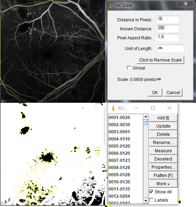
Figure 2.
Comparison of changes in non-perfused retinal area estimated using perifoveal fluorescein angiography. The angiography image is analyzed using ImageJ software (version 1.52a 23/04/2018 by ImageJ, National Institutes of Health, Wayne Rasband, Bethesda, Maryland, USA), which is an automated system used for accurate image analysis (distance in pixels: 16, known distance: 200µm, pixel aspect ratio: 1.0).

