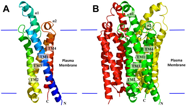
Figure 1.
Structure of the Chlamydomonas reinhardtii LCI1. (A) Ribbon diagram of an LCI1 monomer viewed in the membrane plane. The molecule is colored using a rainbow gradient from the N-terminus (blue) to the C-terminus (red). (B) Ribbon diagram of an LCI1 trimer viewed in the membrane plane. The three protomers are colored green, red and yellow, respectively. The transmembrane segments (TMs) and α-helices (αs) of the front protomer (green) of LCI1 are labeled. The Figure was prepared using PyMOL (http://www.pymol.sourceforge.net).

