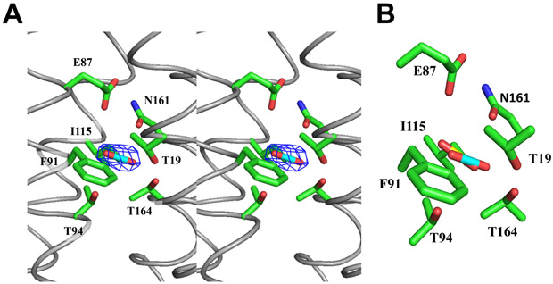
Figure 4.
Putative CO2 binding site. (A) Stereo view of the Fo - Fc electron density map of bound ligand, presumed to be CO2, in LCI1, with the putative bound CO2 ligand and residues putatively involved in CO2 binding shown as a stick model (cyan, carbon; blue, nitrogen) and as green sticks, respectively. The Fo - Fc map is contoured at 3.0 σ (blue mesh). (B) A composite figure showing the locations of the predicted bound CO2 ligand (yellow) and the putative bound CO2 (cyan) identified in the LCI1 crystal structure.

