Fig. 3.
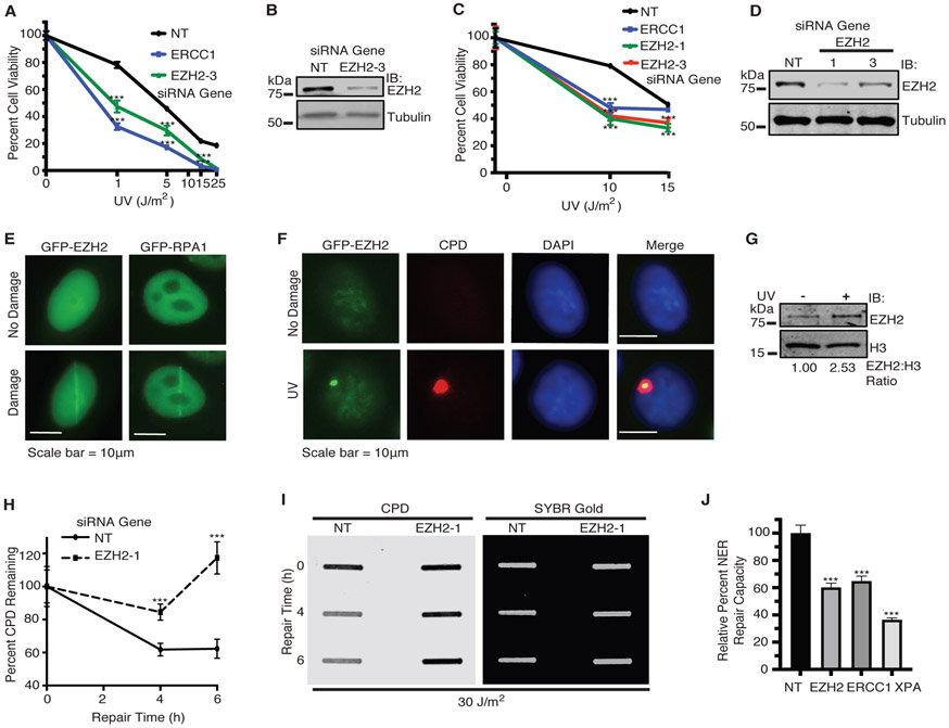
EZH2 localizes to and promotes repair of UV-induced CPD lesions. a H128 and c H146 cells were transfected with siRNAs targeting EZH2, ERCC1, or a NT control. 72 hours after transfection, cells were treated with UV. Cell viability was measured 72 hours after UV irradiation. b and d Western blot analysis of EZH2 expression (for b H128; and d H146 cells) demonstrating knockdown. e HeLa cells were transiently transfected with GFP-EZH2 or GFP-RPA1. 72 hours post-transfection, cells were subjected to laser microirradiation at 365 nm wavelength. Representative images of GFP localization as seen before and seconds after laser microirradiation are shown. Scale bars represent 10 μm. f HeLa cells were transfected with GFP-EZH2 and 24 hours post transfection, cells were UV irradiated at 100 J/m2 through a 5 μm micropore membrane. Cells were fixed and stained with the indicated markers after one hour recovery. Representative images are shown. g HeLa cells were left untreated or UV irradiated at 30 J/m2. Cells were lysed and fractionated to obtain the chromatin soluble fraction by salt extraction followed by 10 minutes of MNase digestion at room temperature. This fraction was run on SDS-PAGE and probed for EZH2 and H3 for loading. h and i Slot blot analysis of the repair of CPD lesions over time in H128 cells in response to 30 J/m2 of UV. h Quantitation of percent of CPD lesions remaining over time. i Representative slot blot. SYBR Gold signal indicates total DNA loaded. (For a, c, and h, mean and standard deviation of three replicas is shown. j NER repair capacity as measured by FM-HCR was quantified in cells transfected with siRNAs targeting EZH2, ERCC1, XPA or NT control. Relative NER repair capacity was normalized to a NT control. *** indicates p<0.001. For g and i-j, representative blots from 3 independent experiments (n=3) is shown).

