
Extended Data Fig. 2 |. identification of mGFP positive cells.
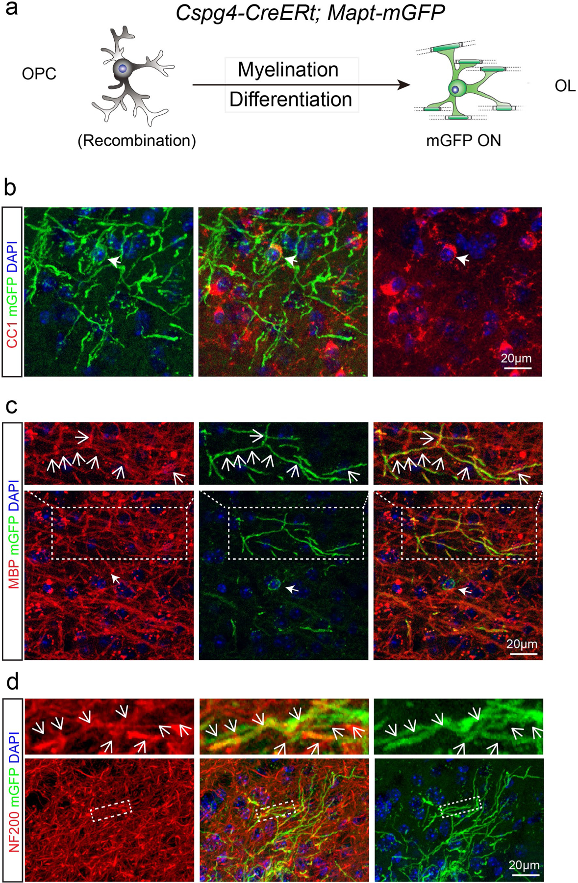
a, Schematic illustration showing mGFP expression in the Cspg4-CreERt; Mapt-mGFP mice; b–d, Immunostaining indicates that mGFP positive cell bodies are CC1 positive (b) and mGFP positive segments are co-localized with MBP (c) and associated with NF200 positive axons (d). These experiments were repeated 3 times independently with similar results, scale bar = 20 μm.