
Figure 3.
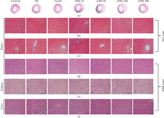
Effect of AMS on histopathology. (a) Transverse section of the heart representing the ventricular diameter. (b, c) Masson's trichrome staining of the heart tissue representing interstitial and perivascular fibrosis, respectively. (d–f) Haematoxylin and eosin stain of the heart, kidney, and liver.