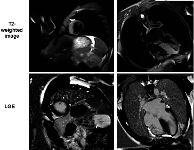
Figure 3.

Cardiac magnetic resonance images of late gadolinium enhancement (LGE) and T2-weighted imaging in patients with hypertrophic cardiomyopathy. The LGE images (lower panel) and T2-weighted images (upper panel) are shown at the identical slice location in the same view. The areas of high T2 signal do not correspond well to those of LGE in patients with HCM.
