
Figure 1.
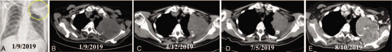
Radiological images of the pulmonary sarcomatoid carcinoma (indicated by arrow) before and after treatment in case 1. A. The X-ray showed a bulky mass located in left upper thorax on admission. B. Further computed tomography revealed the pulmonary tumor invading adjacent ribs. C. The tumor showed PR after 3 months of treatment. D. The tumor was shorter partial remission after 6 months of treatment. E. The lesion was significantly enlarged on the 7-month follow up. CT = computed tomography, PR = partial remission.