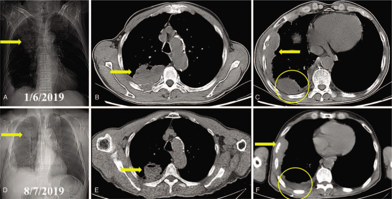
Figure 3.

Radiological images of the thoracic lesions (indicated by arrow and circle) before and after systemic treatment in case 3. A. The X-ray showed a bulky mass located in right upper thorax. B. Computed tomography revealed 1 of the pulmonary tumors invading adjacent ribs. C. The other thoracic tumors with osteolytic rib destruction and thickened pleura were shown. D. X-ray indicated a smaller lesion in right upper thorax. E. The tumor showed partial response after 6 months of treatment. E. The other lesions and osteolytic ribs also revealed partial response. CT = computed tomography, PR = partial response.
