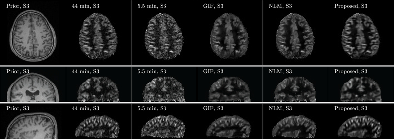
FIGURE 4.
Three orthogonal views of denoising result from Subject 3 (S3). Images of anatomical prior (first column) and averaged perfusion maps from 44 min acquisition (second column), 5.5 min acquisition (third column), 5.5 min acquisition with GIF denoising (fourth column), 5.5 min acquisition with NLM denoising (fifth column), and 5.5 min acquisition with the proposed denoising method (sixth column) are shown. Note that the averaged perfusion maps from 44 min acquisition serve as the ground-truth image.

