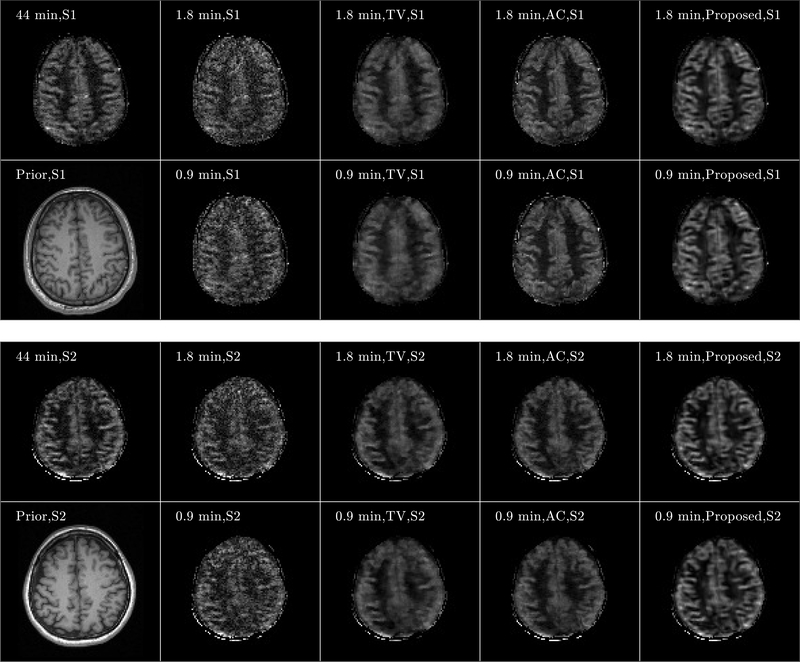
FIGURE 8.
Axial view of image reconstruction results from Subjects 1 (S1) and Subject 2 (S2). Results from S1 and S2 are shown from the top and bottom subfigures, respectively. For each subfigure, images of anatomical prior (1st column, 2nd row) and averaged perfusion maps from 44-min acquisition (1st row, 1st column), SENSE reconstruction for 1.8-min (2nd column, 1st row) and 0.9-min (2nd column, 2nd row) acquisitions, TV penalized reconstruction for 1.8-min (3rd column, 1st row) and 0.9-min (3rd column, 2nd row) acquisitions, anatomically constrained (AC) penalized reconstruction for 1.8-min (4rd column, 1st row) and 0.9-min (4rd column, 2nd row) acquisitions, and reconstruction with the proposed framework for 1.8 min (5th column, 1st row) and 0.9 min (5th column, 2nd row) acquisitions are shown. Note that the averaged perfusion maps from 44-min acquisition serve as the ground-truth image.

