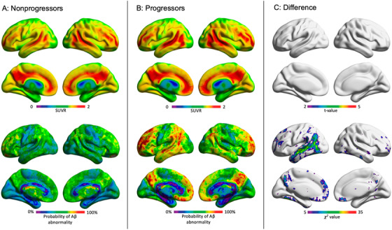
FIGURE 3.

Amyloid beta (Aβ) positive mild cognitive impairment (MCI) progressors were more likely to have Aβ abnormality in the default mode network than nonprogressors. While all Aβ positive MCI individuals (progressors and non‐progressors) demonstrated similar voxel‐wise and global standardized uptake value ratios (SUVRs), they exhibited different patterns of voxel‐wise amyloid positivity. A, Bottom row: amyloid positive MCI individuals who remained stable over 2 years did not exhibit a consistent pattern of regional Aβ positivity. B, In contrast, individuals who progressed to dementia in a 2‐year time frame had higher probability of abnormality in the posterior cingulate/precuneus, lateral temporal, medial and lateral prefrontal cortices. C, Result of voxel‐wise statistical comparisons between Aβ positive MCI progressors versus non‐progressors. Top row: there was no significant difference in voxel‐wise [18F]florbetapir SUVR between progressors and non‐progressors after correcting for multiple comparisons with a false discovery rate of P < .05. Bottom row: Results of voxel‐wise χ2 comparing abnormal voxels (coded as: normal = 0 and abnormal = 1) between Aβ positive MCI progressors versus non‐progressors. Aβ positive MCI individuals who progressed to Alzheimer's disease were significantly more likely to have Aβ abnormality in large clusters in the posterior cingulate cortex/precuneus, medial prefrontal cortex, and lateral temporal cortices. Results are corrected for multiple comparisons with a false discovery rate of P < .05
