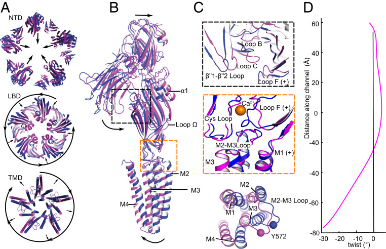
Fig. 5.
Conformational state transitions at the tertiary and quaternary levels. (A) Superposition of DeCLIC structures in the presence (blue) and absence (magenta) of Ca2+, viewed from the periplasmic side, illustrating relative conformational changes in the NTD (Top), LBD (Middle), and TMD (Bottom). Loop regions except the M2–M3 loop are hidden for clarity. Arrows indicate predominant quaternary rearrangements involving radial contraction/expansion or tangential twist. (B) Conformational changes in a single subunit between Ca2+-bound (blue) and -free (magenta) states, aligned by superimposition of the entire pentamer, viewed from the membrane plane. Arrows indicate predominant motions involving contraction of the NTD and LBD, and expansion of the TMD. (C) Details as in B showing remodeling at the NTD2–LBD interface (Top), LBD–TMD interface (Middle), or of a single TMD subunit viewed from the periplasmic side (Bottom). (D) Twist angle (magenta) between Ca2+-bound and -free states in successive z slabs along the linear channel axis. Negative values in the lower channel correspond to a relative clockwise twist of the TMD.

