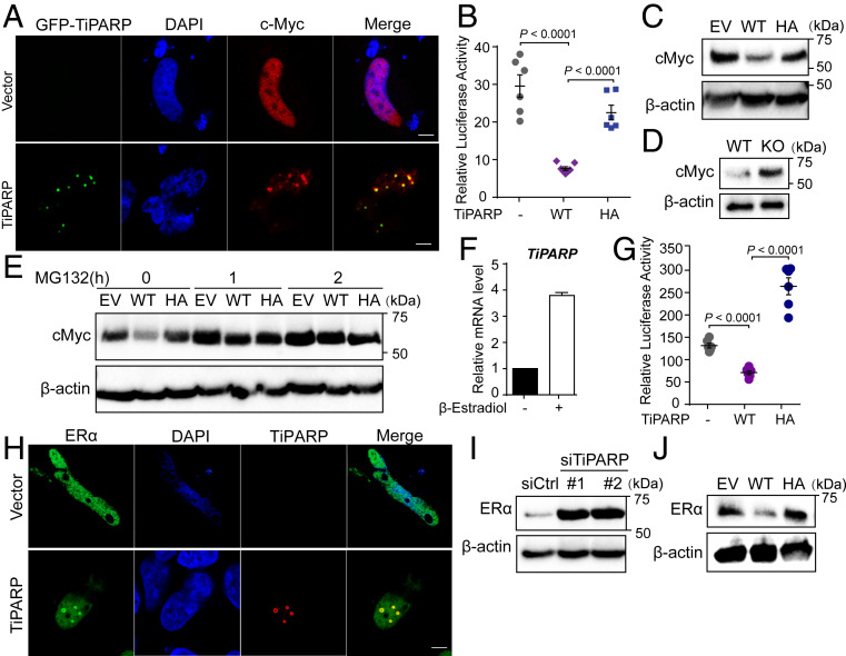
Fig. 6.
TiPARP inhibits ERα and c-MYC. (A) HEK 293T cells were cotransfected with Flag-cMyc, as well as empty vector (EV) or GFP-TiPARP (green). c-Myc was stained with anti-FLAG (red) antibodies. (Scale bar, 5 μm.) (B) Transcriptional activity of c-Myc was measured using a luciferase reporter with c-Myc binding sites. c-Myc was cotransfected with empty vector, TiPARP WT, or H532A mutant (HA) in HEK 293T cells. Data are represented as means ± SEM (n = 6). (C) Western blot analysis of cMyc in HEK 293T cells expressing empty vector (EV), Flag-tagged WT, or H532A inactive mutant (HA) TiPARP. (D) Western blot analysis of cMyc in WT and TiPARP KO HAP-1 cells. (E) Western blot of cMyc in HCT116 cells expressing empty vector (EV), Flag-tagged WT, or H532A mutant of TiPARP and treated with or without MG132. (F) qRT-PCR analysis of TiPARP mRNA expression in MCF-7 cells treated with DMSO or 10 nM β-estradiol for 24 h. Data are represented as means + SD (n = 8). (G) Transactivation of ERα measured using a luciferase reporter containing estrogen response elements (EREs). HEK 293T cells were transfected with ERE-luciferase, Flag-TiPARP, together with HA-tagged ERα. Twelve hours after transfection, cells were treated with 1 μM β-estradiol for 24 h, followed by luciferase measurement. Data are represented as means ± SEM (n = 6). (H) HeLa cells were cotransfected with HA-tagged ERα and empty vector (EV) or Flag-TiPARP. Colocalization was analyzed by immunofluorescence with anti-HA (green) and anti-FLAG (red) antibodies. Nuclei were stained with DAPI (blue). (Scale bar, 5 μm.) (I) Western blot analysis of endogenous ERα in MCF-7 cells transfected with control or TiPARP siRNA. (J) Western blot of endogenous ERα in HCT116 cells expressing empty vector (EV), Flag-tagged WT, or H532A mutant (HA) of TiPARP.

