FIGURE 2.

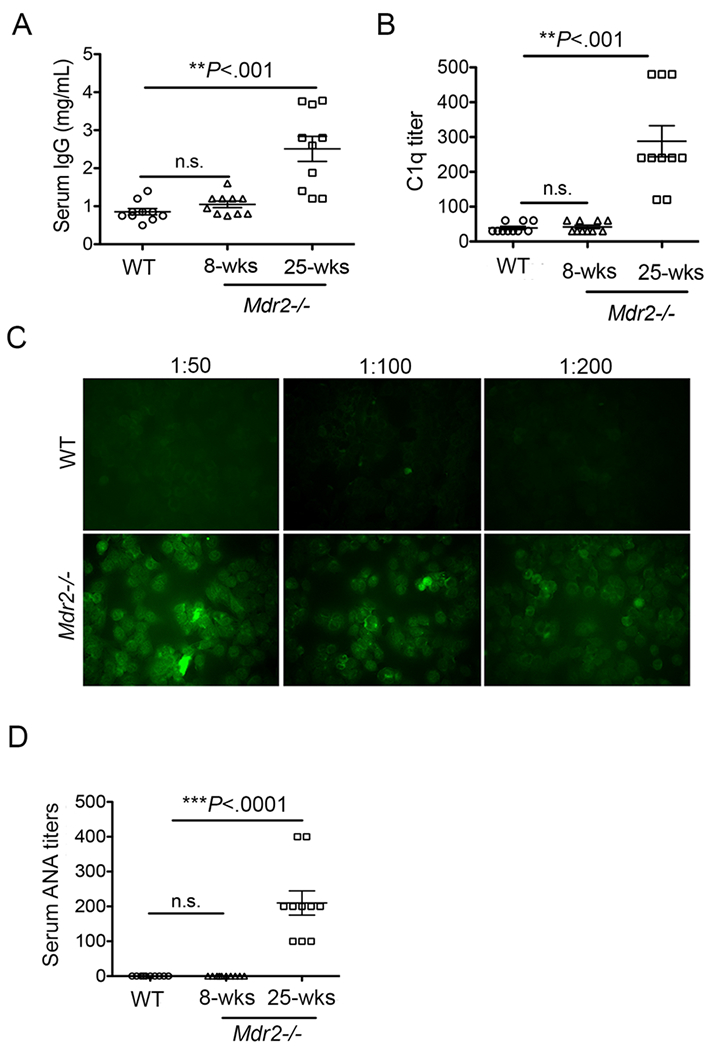
Elevated autoantibodies and immune complexes in Mdr2−/− mice. Serum specimens were collected from 8-weeks old and 25-weeks old Mdr2−/− and WT animals, and processed for detection of (A) total IgG by using a standard IgG ELISA technique, (B) total C1q titers by using a modified ELISA technique, and (C) Representative ANA images by using immunofluorescence staining of Hep-2 substrate are shown. (D) ANA titers are shown in 8-weeks old and 25-weeks old Mdr2−/− mice and WT controls. Data are representative of at least 3 independent experiments (n=3-5 per group).
Statistical significance was determined by a two-tailed Mann-Whitney Test, *P<.05, **P<.005, ***P<.0001. Error bars represent the SEM.
