
FIGURE 7.
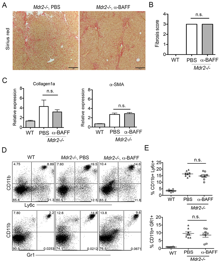
Blockade of BAFF do not attenuate hepatic fibrosis. (A) Mdr2−/− mice were administered anti-BAFF mAb as 250μg in sterile PBS i.p. every 7 days starting at 8 weeks continued until 25 weeks of age. Histologic analysis of representative liver specimens for Sirius red staining (mag. 100×) are shown. (B) Blind fibrosis scoring of Mdr2−/− animals following antibody treatment is shown. (C) Relative gene expression of liver collagen1a and α-SMA by PCR analysis following antibody treatment is shown. (D) Representative FACS plots of intrahepatic myeloid populations monocytes (CD11b+Ly6c++), and neutrophils (CD11b+Gr1+) are shown. (E) Frequencies of myeloid populations monocytes and neutrophils are shown. Statistical significance was determined by a two-tailed Mann-Whitney Test. *P<.05. Error bars represent SEM.