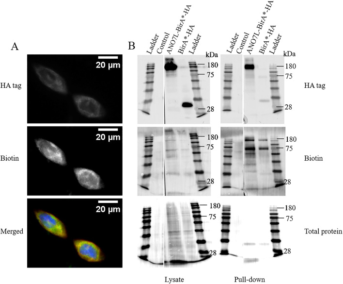
Figure 2.
Biotinylation of proteins in ANO7L-Bir-HA-transfected LNCaP cells. A. Immunohistochemical staining of HA-tagged ANO7L-Bir (red) and biotinylated proteins with Alexa Fluor 488-tagged streptavidin (green) and nuclear staining with DAPI (blue). B. The insoluble cell lysate fraction is on the left side, and streptavidin bead pull-down samples are on the right side. The control lane contains untransfected LNCaP cells treated with biotin. Expression of the fusion proteins was detected with anti-HA, biotinylated proteins were detected with streptavidin-HRP, and total protein were detected with Ponceau S staining. The staining was visualized with Nikon Eclipse Ni-U upright fluorescence microscope (Nikon Instruments, Inc. Shinagawa, Tokyo, Japan).

