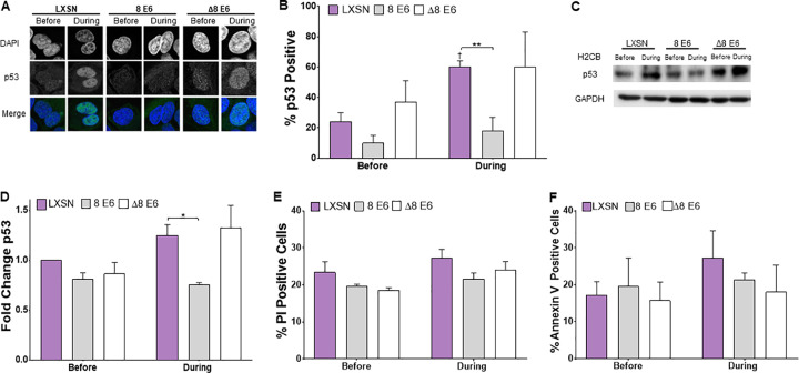
FIG 5.
β-HPV 8E6 attenuates p53 accumulation upon H2CB-induced failed cytokinesis. (A) Representative images of p53 and DAPI staining in cells before and during H2CB treatment. (B) Percent of p53-positive U2OS cells. (C) Representative immunoblot of p53 before and during H2CB exposure. (D) Densitometry of immunoblots described in panel C. GAPDH was used as a loading control. Data were normalized to p53 levels in untreated LXSN cells (set to 1). (E) Percent of propidium iodide-stained HFK cells before and during H2CB exposure. (F) Percent of annexin V-stained HFK cells before and during H2CB treatment. At least 200 cells/line were imaged from three independent experiments. Figures depict mean ± standard error of the mean; n ≥ 3. *, significant difference between indicated samples; †, significant difference relative to before H2CB; one symbol (* or †), P ≤ 0.05; two symbols (** or ††), P ≤ 0.01; three symbols (*** or †††), P ≤ 0.001 (Student's t test). In β-HPV Δ8E6, residues 132 to 136 were deleted.

