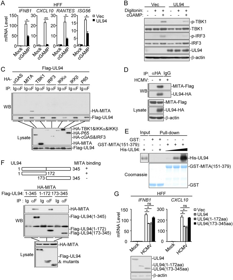
FIG 5.
UL94 targets MITA. (A) UL94 inhibits cGAMP-induced transcription of antiviral genes in HFFs. HFFs stably expressing UL94 (4 × 10⁵) were transfected with cGAMP (0.2 μg/ml) for 3 h before qPCR analysis. (B) UL94 inhibits cGAMP-induced phosphorylation of TBK1 and IRF3 in HFFs. HFFs stably expressing UL94 (4 × 10⁵) were transfected with cGAMP (0.2 μg/ml) for 3 h before immunoblot analyses were performed with the indicated antibodies. (C) Association of UL94 with MITA. HEK293T cells (2 × 10⁶) were transfected with the indicated plasmids for 24 h. Co-immunoprecipitation and immunoblot analyses were performed with the indicated antibodies. (D) Endogenous association of UL94 with MITA following HCMV infection. HFFs stably expressing Flag-tagged MITA (2 × 10⁷) were infected with HCMV-UL94-HA (MOI = 3) for 72 h. Co-immunoprecipitation and immunoblot analyses were performed with the indicated antibodies. (E) UL94 directly binds to MITA. Purified GST-MITA (151 to 379) was bound to glutathione agarose beads and incubated with purified His-UL94 for 3 h. Immunoblot analyses were performed with the indicated antibodies. (F) Domain mapping of the UL94-MITA association. HEK293T cells (2 × 10⁶) were transfected with the indicated UL94 truncation mutants for 24 h before co-immunoprecipitation and immunoblot analyses were performed with the indicated antibodies. (G) Effects of UL94 truncations on HCMV-induced transcription of antiviral genes in HFFs. HFFs stably expressing UL94 or the indicated UL94 truncations (4 × 10⁵) were left uninfected or infected with HCMV (MOI = 1) for 12 h before qPCR analysis. The protein levels of UL94 and its truncations were examined with immunoblot assays. Graphs show means ± SDs, n = 3. *, P < 0.05; **, P < 0.01 (unpaired t test).

