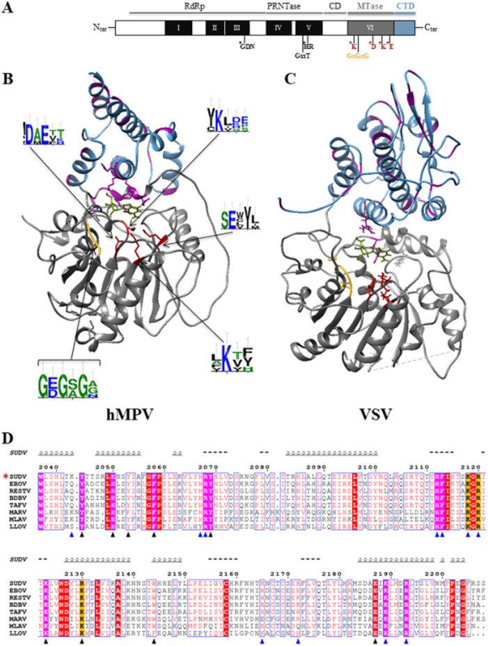
FIG 1.
Bioinformatic analysis of the C-terminal domains (MTase+CTD) of mononegavirus L protein based on hMPV and VSV structures and alignment of the filovirus CTD sequences. (A) Domain organization of the mononegavirus L protein with the six conserved regions (CRI to CRV, black boxes; CRVI, gray box) that contain motifs important for the different activities of the L protein (motifs mapped with asterisks). The Sudan ebolavirus (SUDV) methyltransferase domain (MTase) encompasses amino acids 1693 to 2036, based on the alignment with the vesicular stomatitis virus (VSV) L protein. The SAM-binding site (GxGxG) and the 2′-O catalytic tetrad K-D-K-E are also shown (asterisks). The C-terminal domain (CTD) of SUDV L follows the MTase domain (amino acids 2037 to 2210). (B) X-ray structure of human metapneumovirus (hMPV) MTase (gray) and CTD (blue) domains at a resolution of 2.2 Å (26) (PDB ID, 4UCZ). The residues of the catalytic tetrad and of the SAM-binding site are in red and yellow, respectively. Aromatic and basic residues are in purple. WebLogo (47) was used to illustrate the degree of amino acid conservation based on previously published alignments (34). (C) Structure of VSV MTase (gray) (PDB ID, 5A22) and CTD (blue) (53). The residues involved in the catalytic tetrad and SAM-binding site are in red and yellow, respectively. Aromatic and basic residues are in purple. (D) CTD domains in the following L protein sequences selected from GenBank: EBOV (Zaire ebolavirus, AAG40171.1), SUDV (Sudan ebolavirus, YP_138527.1), TAFV (Tai Forest ebolavirus, ALT19766.1), BDBV (Bundibugyo ebolavirus, AKB09568.1), RESTV (Reston ebolavirus, APA16576.1), MARV (Marburg virus, CAA82542.1), LLOV (Lloviu cuevavirus, YP_004928143.1), and MLAV (Mengla dianlovirus, AZL87829.1). An asterisk indicates the strain under study. The alignment was generated with Seaview and analyzed with ESPript. The numbers on top of the alignment indicate the amino acid positions in the SUDV sequence. Strictly conserved aromatic and basic amino acids are highlighted in purple, and arginine or lysine basic residues are highlighted in orange. Mutations leading to soluble proteins are indicated by blue triangles, and mutations leading to insoluble proteins are indicated by black triangles. Spirals just above the alignment indicate the positions of α helices, based on the secondary structure prediction server (NPS@: PHD secondary structure prediction).

