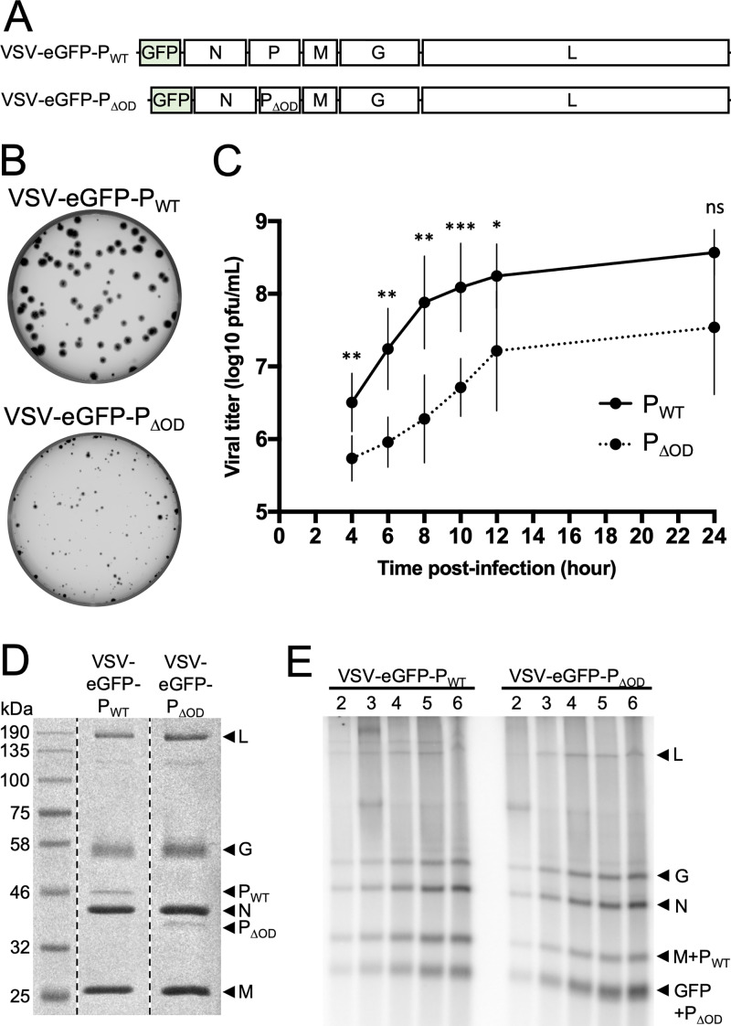
FIG 3.
Characterization of a recombinant VSV expressing PΔOD. (A) Schematic representation of the recombinant virus genomes. (B) Viral spreading as seen by plaque assay on Vero cells infected with VSV-eGFP-PWT or VSV-eGFP-PΔOD. (C) Viral growth kinetic on Vero cells infected with VSV-eGFP-PWT (PWT, black line) or VSV-eGFP-PΔOD (PΔOD, dotted line) at an MOI of 3. Supernatants were harvested and titers determined at 4, 6, 8, 10, 12, and 24 h postinfection. Statistical analysis was performed by a paired t test. * P, < 0.05; ** P, < 0.005; *** P, < 0.0005; ns, nonsignificant. (D) Analysis of virion protein content. Gradient-purified virions (108 PFU) were denaturated by SDS and heat and analyzed by SDS-PAGE and Coomassie staining. (E) In phosphate-free media supplemented with radioactive [32P]orthophosphate, BSR-T7 cells were treated with 10 μg/ml actinomycin D and 100 μg/ml cycloheximide and infected at an MOI of 100 with VSV-eGFP-PWT or VSV-eGFP-PΔOD. RNA was extracted at 2, 3, 4, 5, and 6 h postinfection and analyzed on a 1.75% acid-agarose gel containing 6 M urea. Representative experiment (n = 4).

