FIG 3.
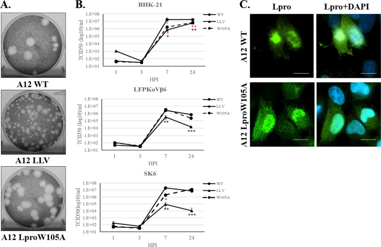
Plaque phenotype and viral growth kinetics. (A) Plaque phenotypes for FMDV A12 WT, A12 LLV, and A12 LproW105A were evaluated in BHK-21 cells. Cell monolayers were infected for 48 h in semisolid medium, followed by staining with crystal violet. (B) Kinetics of growth in multiple cell lines; BHK-21, LFPKαVβ6, and SK6 cells were infected at an MOI of 5 with WT, LLV, or LproW105A FMDV, and at the indicated times, virus titer was measured in BHK-21 cells. FMDV yield was determined by the endpoint dilution method on BHK-21 cells 24 h after infection. Titers are expressed as log10 50% tissue culture infective dose (TCID50) per milliliter. The values are presented as the mean ± standard deviation of three independent experiments. (C) Fluorescent microscopy images from LFPKαVβ6 cells infected with WT or LproW105A FMDV at an MOI of 10 and fixed at 4 h postinfection. Samples were stained with anti-Lpro (green) and with nuclear stain 4′,6-diamidino-2-phenylindole (DAPI; blue). Scale bar represents 10 μm. Statistical analysis was performed using Student’s t test. *, P < 0.05; **, P < 0.005; ***, P < 0.001; black asterisks represent statistical significance between LLV and WT; red asterisks represent statistical significance between W105A and WT.

