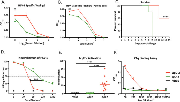
FIG 4.
Differences in passive protection reflect distinct antibody function. (A) Immune serum was assayed for total HSV-specific IgG by ELISA. Results are presented as optical densitometry (OD) units at indicated serum dilution with mean ± standard error of the mean (SEM) (n = 5 mice per group); **, P < 0.01, linear regression. (B) Immune serum from mice with similar HSV-specific IgG titers were pooled, and the new pools were retested in the HSV ELISA (ns, no significant difference in curves by linear regression). (C) Passive transfer studies were repeated with new pools of immune serum containing similar total HSV-specific IgG (n = 5 mice per group). Survival curves were compared by Gehan-Breslow-Wilcoxon test; ****, P < 0.0001 compared to VD60 or adjuvanted rgD-2. (D) Neutralization of viral infection was determined by plaque assay with indicated serum dilution and is shown as percent reduction in PFU relative to control serum. The horizontal line at 50% indicates the dilution of serum that inhibits 50% of viral plaques (neutralization titer). The figure is the average obtained from a representative experiment with 3 individual mice per group (***, P < 0.001 unpaired t test with Welch’s correction area under curve). (E) ADCC was assayed using the murine FcγRIV activation assay with 1:5 dilution of immune serum; results are fold induction relative to controls and data shown as scatterplots (n = 10 mice per group). ****, P < 0.0001 ANOVA with Tukey’s multiple comparison. (F) C1q binding of immune serum was assayed by ELISA at the indicated dilutions, and results are shown as OD mean ± SEM (n = 4 mice per group). *, P < 0.0001 area under curve was compared by unpaired t test with Welch’s correction.

