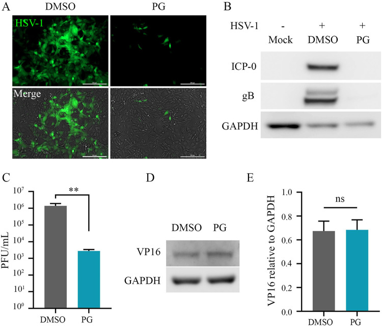
FIG 3.
Prophylactic treatment with prodigiosin inhibits HSV-1. (A to C) HSV-1 (MOI, 0.1) was incubated with PG (2.5 μM), and after 1 h of incubation, the suspension was added over HCE cells and was replaced by DMEM (with 10% FBS and 1% penicillin-streptomycin). DMSO (0.1%) served as a vehicle control. The assay was terminated at 24 hpi, and the cells were collected to analyze HSV-1 infection. (A) Representative micrographs of HCE cells infected with HSV-1 KOS (GFP tagged) at 24 hpi (scale bar, 200 μm). (B) Representative immunoblots for HSV-1 protein ICP0, gB, and GAPDH extracted from HCE cells at 24 hpi. (C) Graph showing mature virus particles produced in the control and treatment groups. (D) Immunoblot of HSV-1 VP16 tegument protein after infecting HCE cells with HSV-1 strain KOS at an MOI of 10. (E) Quantification of HSV-1 VP16 protein, depicting virus entry in HCE cells.

