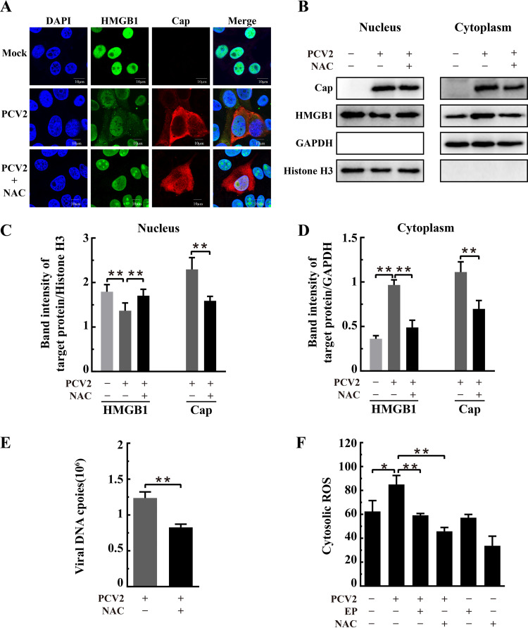
FIG 10.
N-Acetylcysteine inhibited PCV2-induced HMGB1 translocation from nuclei to cytosol and repressed PCV2 replication. PK-15 cells were mock infected or infected with PCV2 (MOI = 1) for 12 h and then treated with 10 mM N-acetylcysteine (NAC). The cell samples were harvested at 36 hpi. (A) Confocal imaging of HMGB1 distribution in PCV2-infected and NAC-treated cells after the cells were fixed and immunostained for HMGB1 (green) and Cap (red). Nuclei were stained with DAPI (blue). Bars, 10 μm. (B) Blotting of HMGB1 and PCV2 Cap in the nuclear and cytoplasmic extracts of PCV2-infected cells with or without NAC treatment. Histone H3 and GAPDH were used as internal controls for the nuclear and cytoplasmic fractions, respectively. The figure is representative of three independent experiments. The ratios of band intensities of HMGB1 or PCV2 Cap to histone H3 (as shown in panel B, left) in the nuclear fraction (C) or to GAPDH (as shown in panel B, right) in the cytoplasmic fraction (D). (E) Effect of NAC on PCV2 genomic DNA replication by qPCR using DNA extracted from lysates of PCV2-infected cells treated with NAC. (F) Cytosolic ROS levels in PCV2-infected cells with or without treatment by NAC or ethyl pyruvate (EP) as measured by flow cytometry after probing with DCFH-DA. Bar charts in panels C, D, E, and F show means ± SDs from three independent experiments. *, P < 0.05; **, P < 0.01.

