Figure 2.
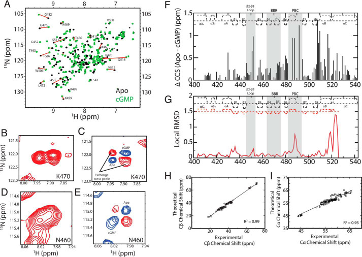
The apo versus cGMP-bound comparative NMR analyses of PfD reveal pervasive allosteric perturbations. A, overlay of 15N-1H HSQC spectra of apo (black) and cGMP-bound (green) PfD samples with representative assignments. Nz-exchange (B and D) and Nz-exchange difference experiments (C and E) utilized to transfer the assignments from the cGMP-bound to the apo spectra. F, CCS differences between the apo and cGMP-bound samples. The secondary structure of PfD is depicted at the top of the plot. The cGMP-binding regions BBR and PBC as well as the adjacent β2–3 loop are highlighted in gray background. G, local apo (PDB code 4OFF) versus cGMP-bound (PDB code 4OFG) root mean square deviation for PfD. The secondary structures obtained from the apo and cGMP-bound crystals are reported as red and black dashed lines, respectively. H, Cβ chemical shift values of cGMP-bound PfD obtained from NMR triple-resonance experiments versus the theoretical Cβ chemical shift values predicted form the structure (PDB code 4OFG) using the ShiftX software (54). I, similar to H but for the Cα chemical shifts.

