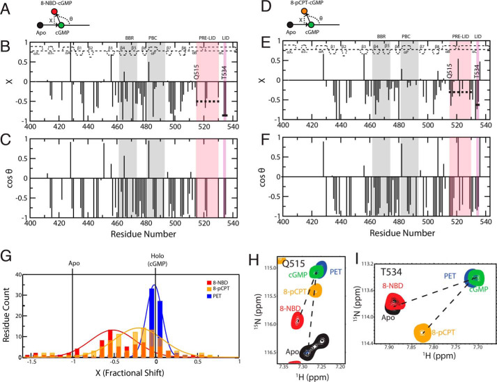
Figure 4.
CHESPA of cGMP analog–bound states reveals a third conformer with a disengaged lid. CHESPA vector scheme, fractional shift (X) and cos(θ) values plotted against residue number for 8-NBD-cGMP (A–C) and for 8-pCPT-cGMP (D–F). The secondary structure of cGMP-bound PfD is depicted at the top of each plot. Pre-lid and lid motifs are highlighted in pink and purple background, respectively. The PBC and BBR are highlighted in gray background. The average 〈X〉 values for residues common in both 8-NBD-cGMP–bound and 8-pCPT-cGMP–bound sample are indicated with a dashed and a solid line for the pre-lid and the lid motifs, respectively. G, distribution of X values for 8-NBD–, 8-pCPT–, and PET-cGMP–bound samples. H and I, HSQC overlay expansions for representative residues in the pre-lid and lid regions of the apo (black) and the 8-NBD-cGMP (red), 8-pCPT-cGMP (orange), PET-cGMP (blue), and cGMP (green) bound samples.

