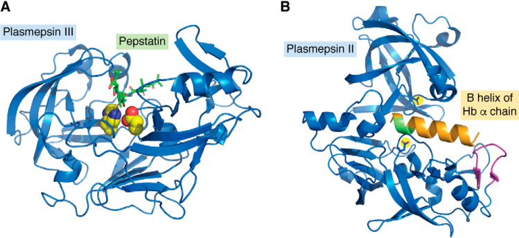
Figure 4.
A, crystal structure of PM III complexed with pepstatin. Shown is a ribbon structure (blue) with Asp215 and His32 highlighted in yellow with red and blue heteroatoms. Pepstatin is in green with red oxygens. The figure was constructed from PDB entry 3FNT. B, crystal structure of PM II (blue) with the B helix of the hemoglobin α chain modeled in orange. The α33–34 cleavage site is green; the helix-interacting loop is magenta; from PDB entry 1PSE. Created using PyMOL Molecular Graphics System, Version 2.3.

