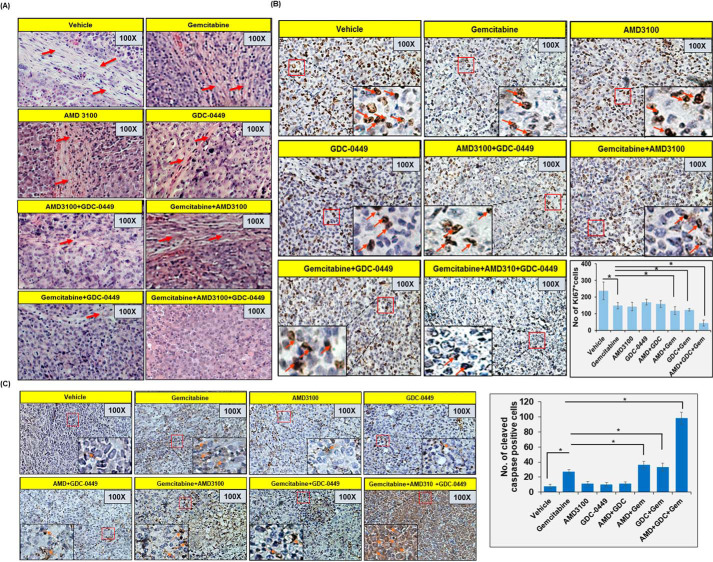
Figure 6.
Immunohistochemistry analysis in pancreatic tumors. A, tissue sections of orthotopically developed pancreatic tumors were deparaffinized, rehydrated, and stained with H&E to study their histopathological characteristics. Desmoplastic region is depicted with red arrows. B and C, tumor sections were also stained with Ki67 (B) and cleaved caspase-3 (C). Representative images show predominantly nuclear and cytoplasmic staining of Ki67 and caspase-3, respectively. All images have been taken at 100 × and 400 × (inset) magnification. Bars in the graphs represent average number of stained cells from five random fields or both ± S.D.; *, p < 0.05.

