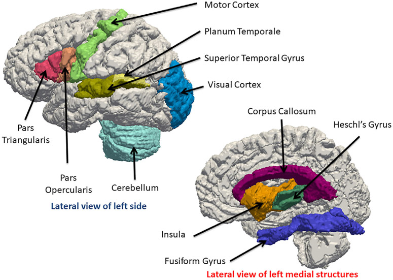
FIGURE 3.
3D visualization of gray matter and white matter structures found to be different in people with hearing loss based on Table 1. Please refer to Figure 2 for the possible roles these structures play in the auditory pathway. Upper left shows the lateral view of the left side of the JHU-MNI-SS brain (Oishi et al., 2009); lower right shows the lateral view of the left medial structures adjacent to the mid-sagittal plane of the right hemi-brain. The cortical structures (Pars Triangularis, Pars Opercularis, Motor Cortex, Superior Temporal Gyrus, Planum Temporale, Visual Cortex and Cerebellum, Heschl's Gyrus, Insula, Fusiform Gyrus) and one white matter structure (corpus callosum) were obtained from the JHU-MNI-SS labels and triangulated. CAWorks (www.cis.jhu.edu/software/caworks) was used for visualization

