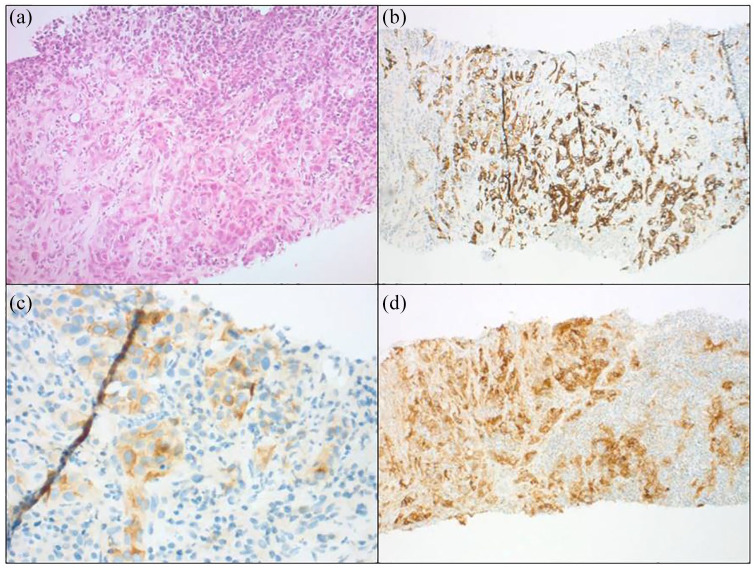
Figure 1.
Moderately differentiated tumour with focal glandular pattern, associated with a dense peritumoural lymphocyte-rich stroma (hemtoxylene eosin and safran × 100). (b) Diffuse expression of cytokeratine 7. (c) Focal positivity with glypican 3 antibody. (d) Strong and diffuse staining for programmed death-ligand 1.

