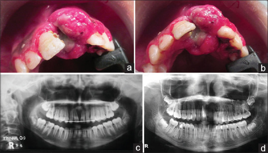
Figure 1.

(a and b) Show a diffuse bright red lesion with a lobulated surface is noted in the labial and palatal aspect extending from 11 to 23 with ill-defined margins; (c) In the initial presentation, the orthopantomography shows a well-defined radiolucency in the interdental region of 11, 21; (d) In the second recurrence, the orthopantomography shows ill-defined radiolucency in the interradicular area extending into the alveolus and palatal region from 11 to 22
