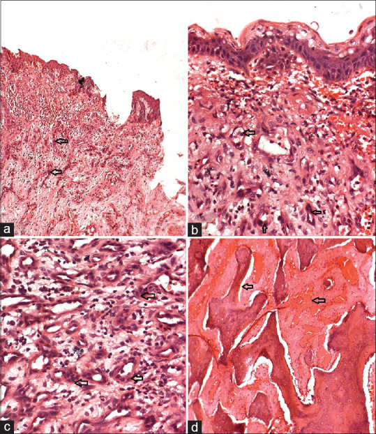
Figure 3.

Second recurrence- (a-c) show the discontinuous oral epithelium with the adjacent stroma showing numerous capillaries lined with plump endothelial cell proliferation and lymphocytic infiltration; (a: H and E stains, low-magnification view ×10; b and c: H and E, high-magnification views ×40); (d) The decalcified section of the bony trabeculae with dilated blood vessels. (H and E, high-magnification views ×40)
