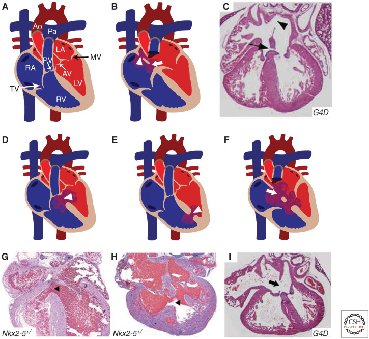
Figure 1.
Murine models of cardiac septation defects: (A) Illustration depicts a cross-sectional view of a normal and mature four-chambered heart. (B) Location of primum (arrowhead) and secundum (arrow) atrial septal defect (ASD) in human heart diagram, which has been recapitulated in Gata4Δex2/WT (G4D) mouse model (C). (D,E) Schematics display similar features of perimembranous and muscular ventricular septal defects (VSDs) (arrowhead) seen in human congenital heart disease (CHD) patients, which were also detected in Nkx2-5+/− mice (G,H). (F) The diagram represents atrioventricular septal defect (AVSD) phenotype with single valve (arrow) noted in humans, resembling the G4D murine model (I). (RA) right atrium, (RV) right ventricle, (LA) left atrium, (LV) left ventricle, (MV) mitral valve, (AV) aortic valve, (PV) pulmonary valve, (TV) tricuspid valve, (Ao) aorta, (Pa) pulmonary artery. (Panels C and I are reprinted from Rajagopal et al. 2007, with permission, from Elsevier © 2007. Panels G and H are reprinted from Winston et al. 2010, with permission, from Circulation © 2010.)

