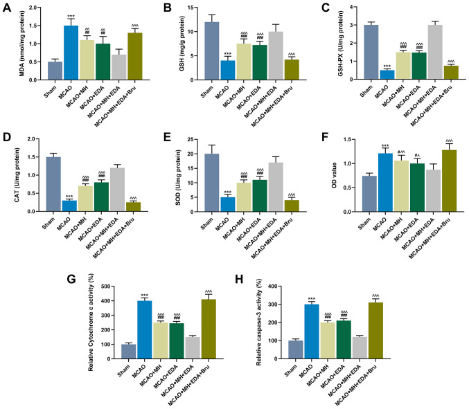
Figure 3.
Effects of MH, EDA and Bru on the levels of oxidative stress indicators, neuronal viability, cytotoxicity and caspase-3 activity. (A-E) The levels of MDA, GSH, GSH-PX, CAT and SOD in rat ischemic hemispheres tissues were determined by colorimetric methods; n=10 mice per group. (F) The viability of neurons from the CA1 region of hippocampi was determined by the MTT assay. (G and H) Cytochrome c and caspase-3 in the mitochondrial fractions from each group was detected by ELISA. ***P<0.001 vs. sham. #P<0.1, ##P<0.01 and ###P<0.001 vs. MCAO. ^P<0.1, ^^P<0.01 and ^^^P<0.001 vs. MCAO + MH + EDA. MCAO, middle cerebral artery occlusion; MH, mild hypothermia; EDA, edaravone; Bru, brusatol; MDA, malondialdehyde; GSH, glutathione; GSH-PX, glutathione peroxidase; CAT, catalase; SOD, superoxide dismutase.

