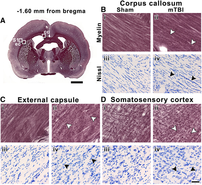
Figure 7.
Representative whole-brain myelin-stained section of a sham-operated animal at −1.60 mm from bregma (A). White squares in panel A indicate the location of high-magnification photomicrographs of myelin-stained sections and Nissl-stained sections of a sham-operated (i and iii) and mTBI animal (ii and iv) in the corpus callosum (B), external capsule (C), and somatosensory cortex (D). The same animals are shown in both stainings. White arrowheads indicate axonal damage, and black arrowheads indicate gliosis shown by increased cellularity in Nissl-staining sections. cc, corpus callosum; ec, external capsule; S1, somatosensory cortex. Scale bars: 2 mm (A) and 50 μm (B–D).

